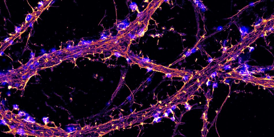
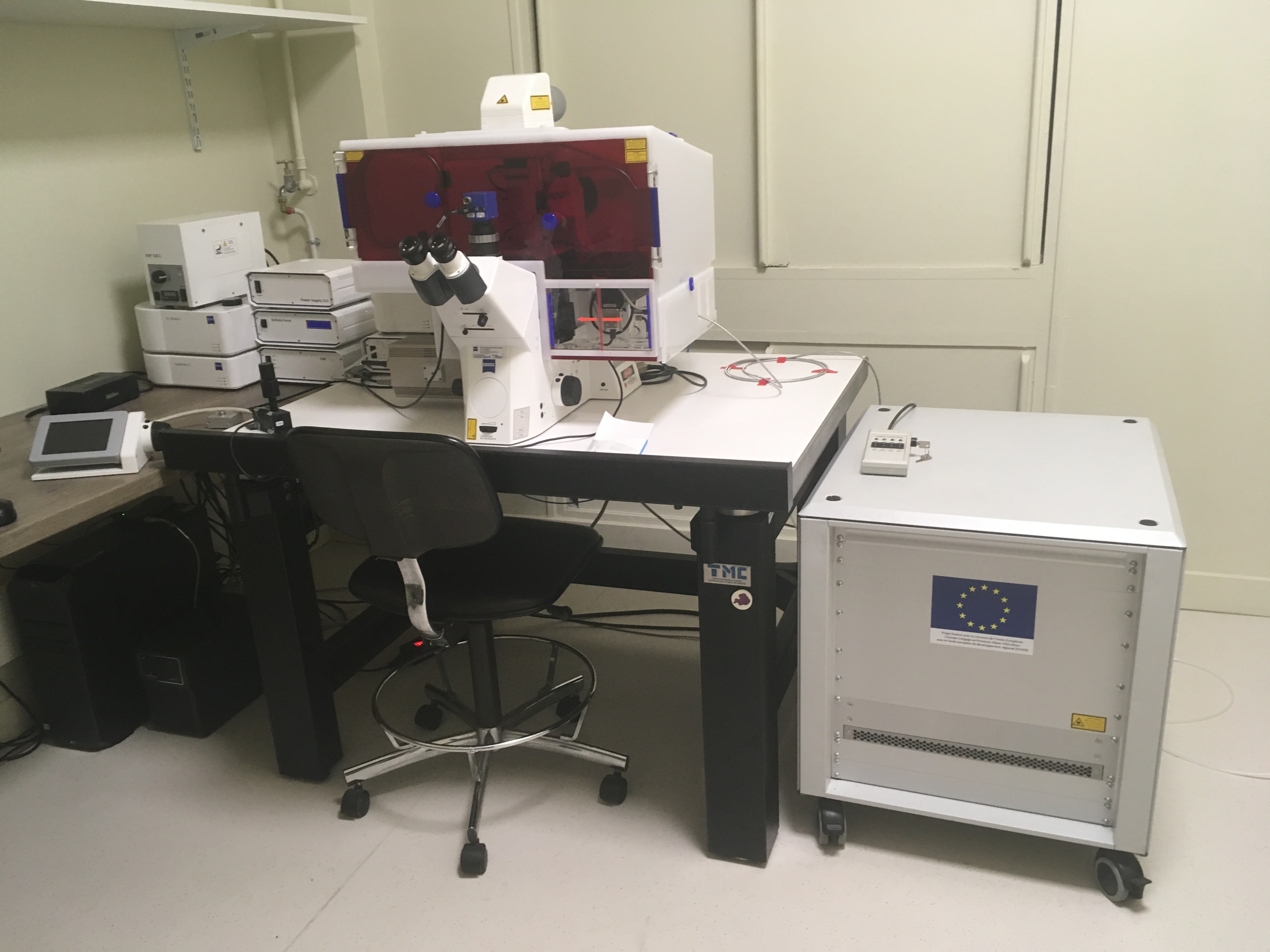
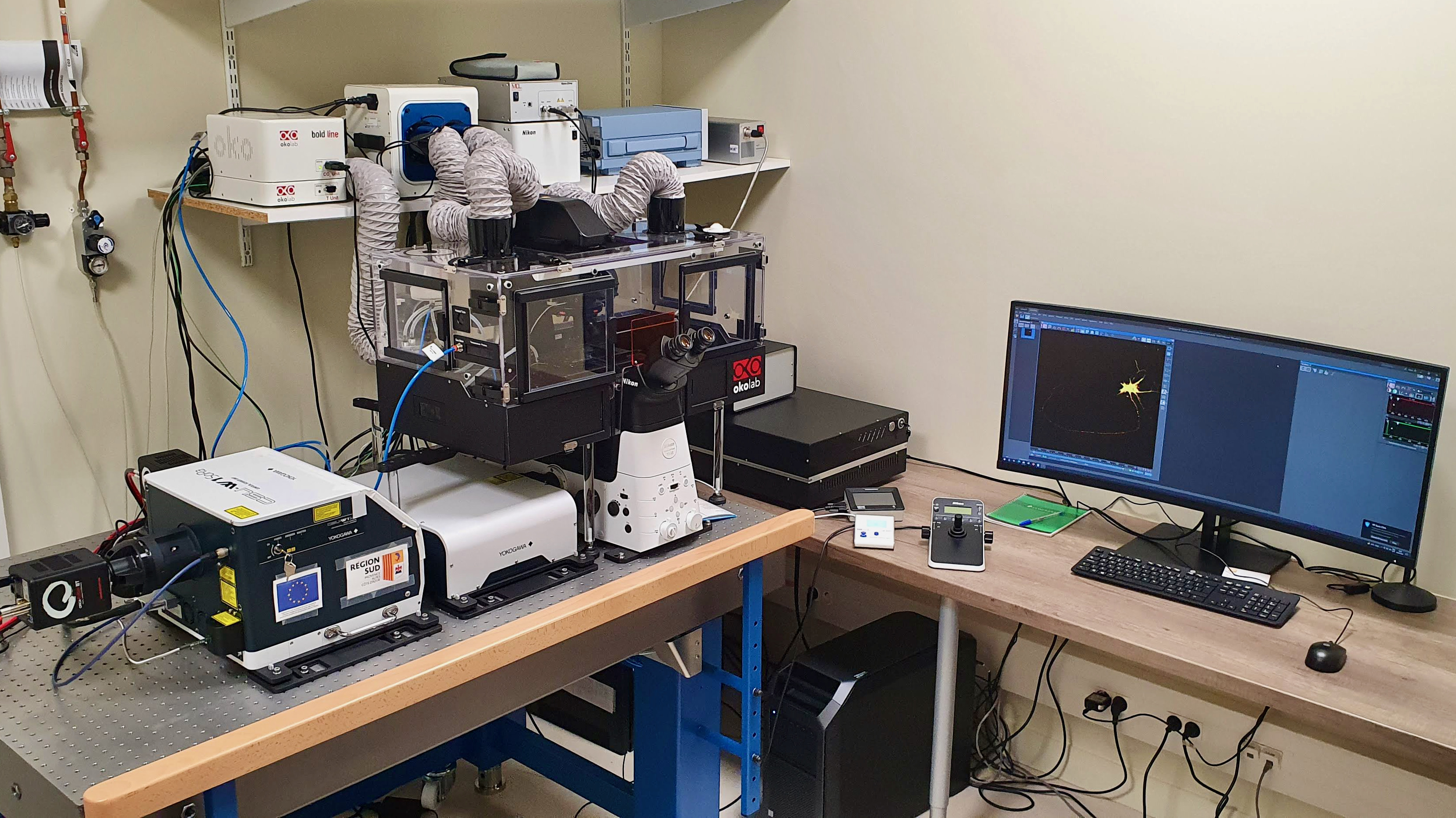
The Neuro-Cellular Imaging Service (NCIS) is part of the Photonic Imaging NeuroTimone (PImaNT) facility. It offers access to optical microscopes for neuroscientists interested in imaging molecular and cellular processes toward better understanding of the functions and dysfunctions of the nervous system. In addition to classical widefield and confocal microscopes, the NCIS provides more advanced techniques such as live-cell imaging, TIRF and super-resolution microscopy (STORM). NCIS staff also offers technical advice, help and training to taylor the imaging and analysis to your scientific question. The NCIS is located in Medicine School building on the Timone campus. Microscopes are accessible to researchers for a fee after initial training on the chosen instrument.
Nikon and the Neurophysiopathology Institute at Aix-Marseille University have partnered to create the Nikon center of Excellence for Neuro-NanoImaging within its cellular imaging facility, focusing on how the latest super-resolutive technique can help in understanding brain cells and their dysfunctions.
For trained users, the reservation of microscopes is available on the site: https://reservations.inp.univ-amu.fr/spip.php?page=intranet_plateformes
If you wish to be trained on a system, technical advice in imaging and analysis, you can contact the engineer in charge of the imaging facility: Laure Fourel
Contact:
Academic Director
Christophe Leterrier, PhD
christophe.leterrier@univ-amu.fr
Facility Manager
Laure Fourel, PhD
Equipements:
WIDEFIELD MICROSCOPY
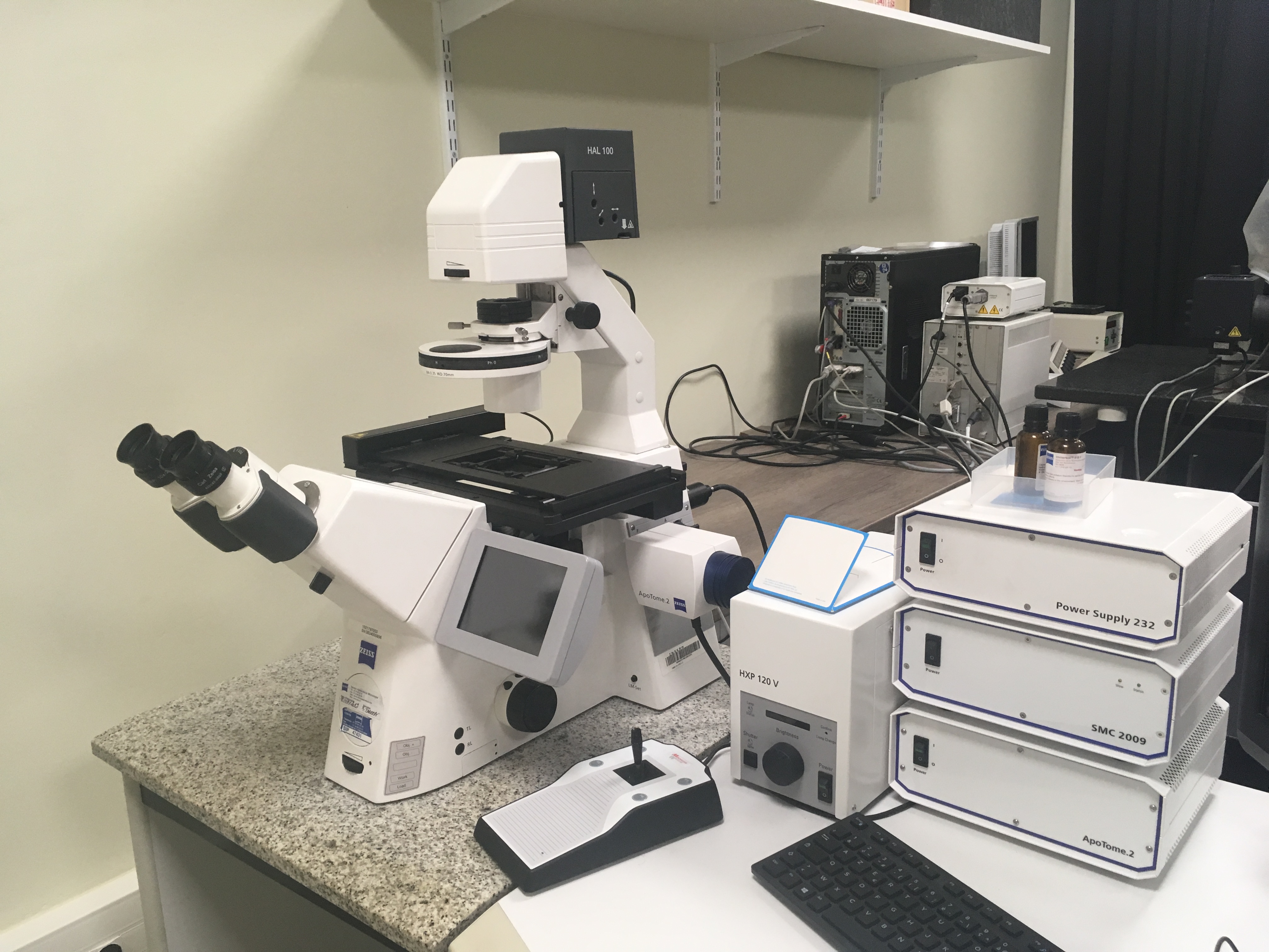
Microscope Zeiss Apotome (salle 133.2)
CONFOCAL MICROSCOPY
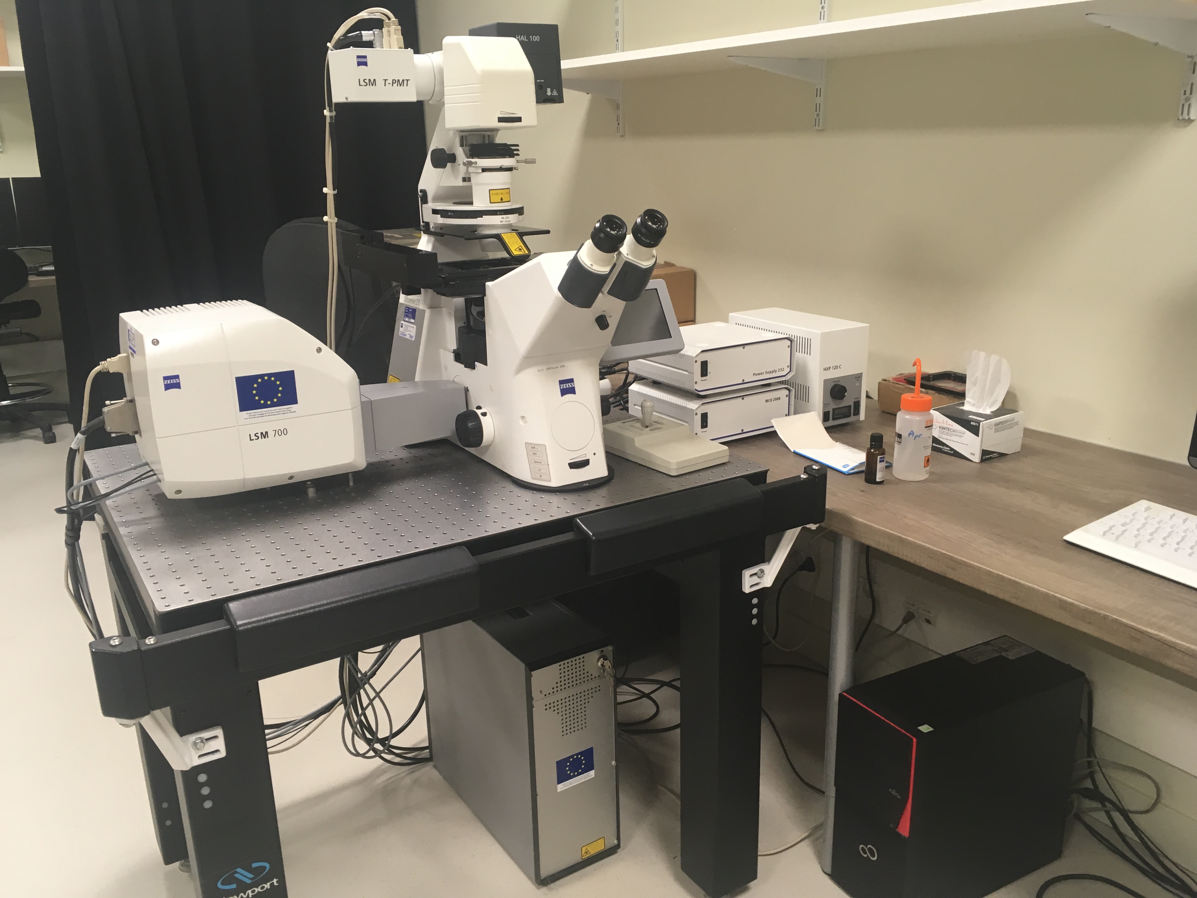
Microscope Zeiss LSM700 (salle 133.2)
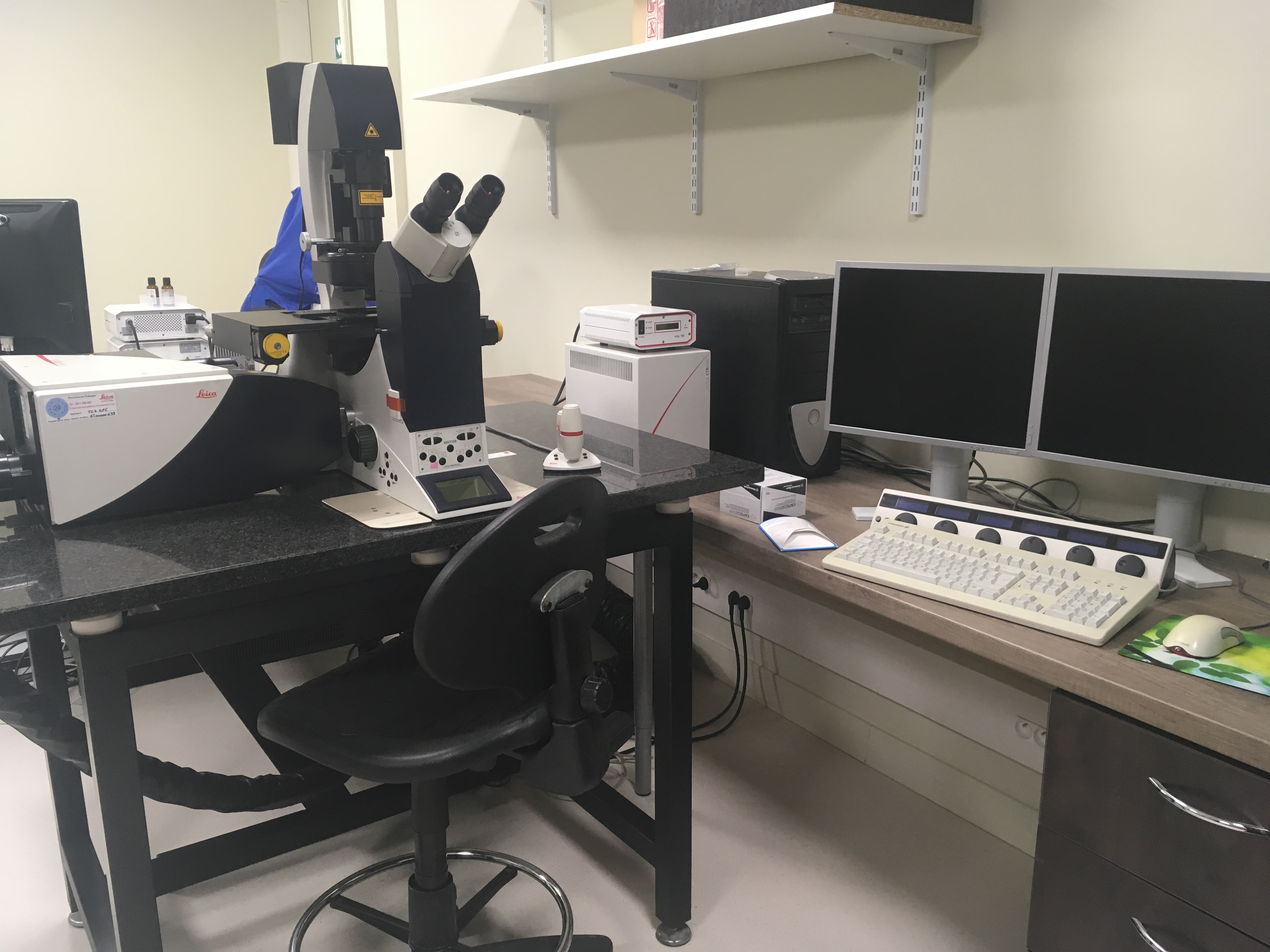
Microscope Leica SP5 (salle 133.2)
VIDEOMICROSCOPY
Microscope Zeiss TIRF (salle 133.1)
Microscope Nikon Spining Disk SoRa (salle 133.2)
SUPER-RESOLUTION MICROSCOPY
Microscope Nikon STORM (salle 133.2)
Microscope Nikon STORM2 (salle 133.3)
Microscope Nikon SIM (salle 133.3)
HIGH CONTENT MICROSCOPY
Perkin Elmer OPERETTA CLS (salle 133.2)
PROCESSING STATION
Nikon processing station (salle 57)


![]()


